What is an MRI of the Extremities?
 Magnetic resonance imaging (MRI) is a noninvasive medical test that helps physicians diagnose and treat medical conditions. MRI uses a powerful magnetic field, radio frequency pulses and a computer to produce detailed pictures of organs, soft tissues, bone and virtually all other internal body structures. The images can then be examined on a computer monitor, transmitted electronically, printed or copied to a CD. MRI does not use ionizing radiation (x-rays). Detailed MRI images allow physicians to evaluate various parts of the body and determine the presence of certain diseases.
MRI imaging is usually the best choice for examining the following:
- Body’s major joints.
- Spine for disk disease.
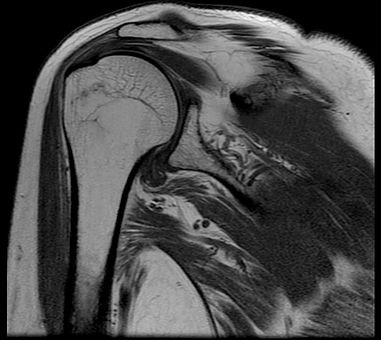
- Soft tissues (muscles, tendons and ligaments) and bones of the extremities.
MRI imaging is typically performed to diagnose or evaluate:
- Degenerative joint disorders such as arthritis, meniscal tears (knee) or labral tears (shoulder and hip)
- Fractures (in selected patients)
- Joint abnormalities due to trauma (such as tears of ligaments and tendons)
- Spinal disk abnormalities (such as a herniated disk)
- The integrity of the spinal cord after trauma
- Sports-related injuries and work-related disorders caused by repeated strain, vibration or forceful impact
- Infections (such as osteomyelitis)
- Tumors (primary tumors and metastases) involving soft tissues around the joints and extremities (such as muscle, bones and joints)
- Pain, swelling or bleeding in the tissues in and around the joints and extremities
- Congenital malformations of the extremities in children and infants
- Development abnormalities of the extremities in children and infants
- Congenital and idiopathic (developing during adolescence) scoliosis prior to surgery
- Tethered spinal cord (abnormal stretching in the spinal cord) in infants and children
*Information courtesy of Radiologyinfo.org.